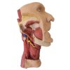
Erler zimmer deep face, infratemporal fossa
Features and Benefits
- In this 3D printed specimen of a midsagittally-sectioned right face and neck, the ramus, coronoid process and head of the mandible have been removed to expose the deep part of the infratemporal fossa.
- The pterygoid muscles have also been removed to expose the lateral pteygoid plate and posterior surface of the maxilla.
- The buccinator has been retianed and can be seen originating from the external aspect of the maxilla, the pterygomandibular raphe and the external aspect of the (edentulous) mandible.
Warranty Information
- 1-Year Warranty


